
When a patient with a suspected stroke reaches hospital, the team has to answer three questions quickly: Is this a stroke? Is it a blockage or a bleed? Can a treatment that opens the artery still help? Tests in the first hour are aimed at these questions. Other tests, done over the next few days, look for the cause so that another stroke can be prevented.
In the emergency department
History and examination
The doctor asks when symptoms started (or when the person was last normal), what medicines they take and about past illnesses. A quick neurological examination checks consciousness, speech, vision, face, arm and leg strength, sensation and coordination. Many hospitals record this as a stroke severity score, which helps guide treatment.
Blood sugar, straight away
Very low blood sugar can cause weakness and confusion that look exactly like a stroke, and is easily corrected. It is checked with a finger-prick test before anything else.
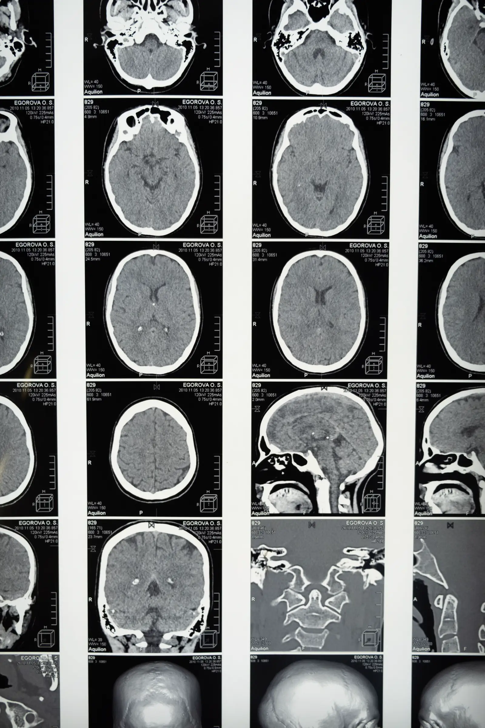
Plain CT scan of the brain
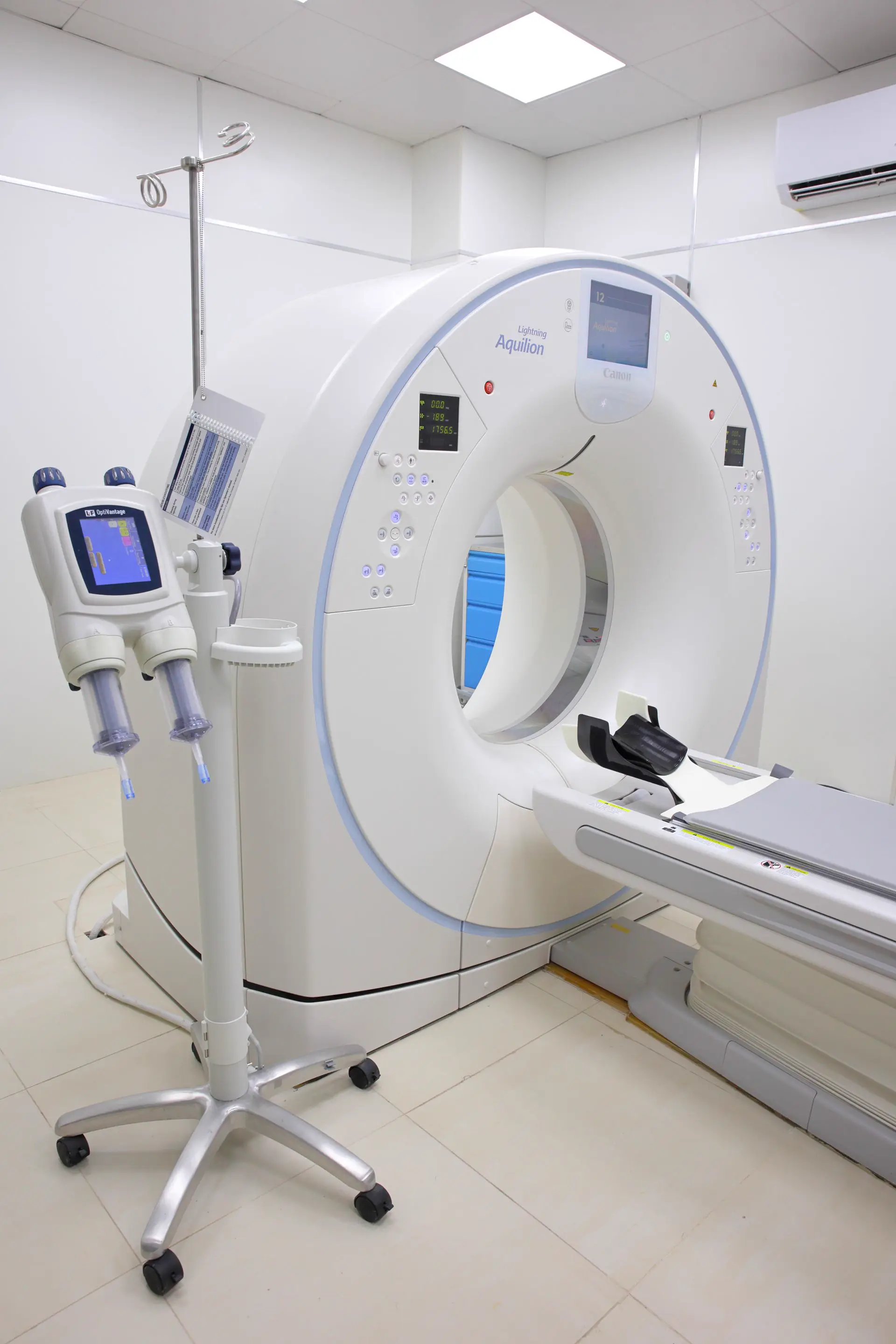
This is the most important first test. It takes a few minutes and shows whether there is bleeding. If there is no bleed and the patient is within the time window, clot-dissolving treatment can be considered. A CT done very early may look normal in an ischaemic stroke; that is expected and does not mean there is no stroke.
CT angiography (CTA) and CT perfusion
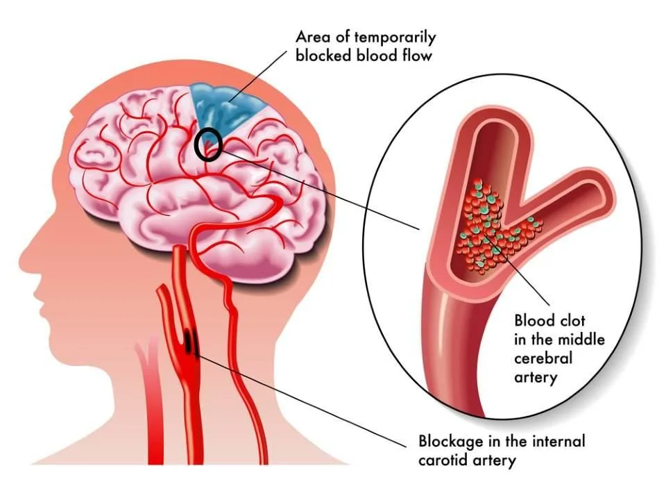
With dye injected into an arm vein, CTA shows the arteries of the neck and brain and whether a large artery is blocked. A large-artery blockage may be treated by removing the clot (mechanical thrombectomy). CT perfusion shows how much brain is already damaged and how much is at risk but still saveable, which helps decide treatment for patients who arrive later.
Blood tests
- Complete blood count, including platelets
- Clotting tests (PT/INR), especially for people on blood thinners
- Kidney function, electrolytes and liver function
- Cholesterol, HbA1c and other tests later, to look at risk factors
ECG
An ECG looks for atrial fibrillation, a recent heart attack or other heart problems that can send clots to the brain.
Over the following days
- MRI brain: more detailed than CT. Diffusion-weighted MRI shows even small, fresh areas of stroke, and can confirm a stroke when CT is unclear, including in the brainstem.
- MR angiography: pictures of the arteries without X-rays.
- Carotid Doppler ultrasound: checks the neck arteries for narrowing caused by plaque.
- Echocardiogram: an ultrasound of the heart to look for clots, valve problems or a hole between the heart chambers.
- Holter or longer heart-rhythm monitoring: to find atrial fibrillation that comes and goes.
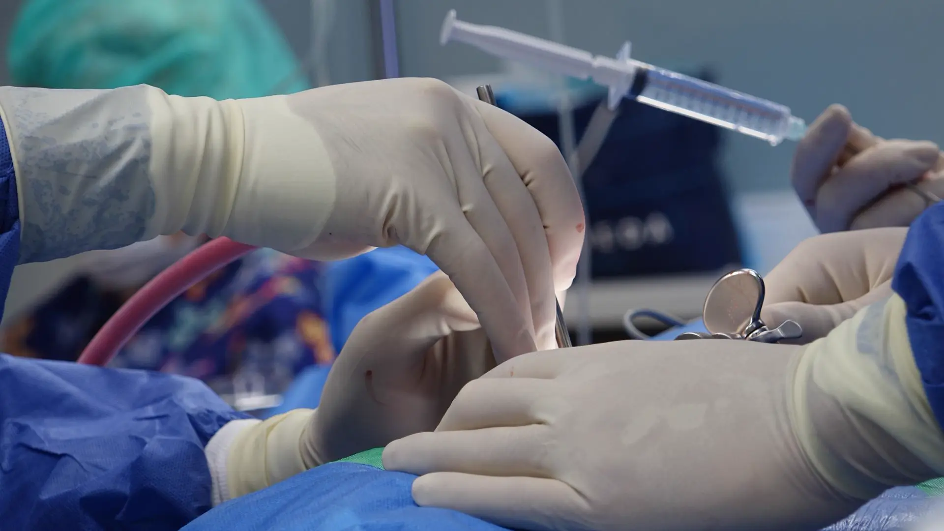
- Digital subtraction angiography (DSA): a catheter test that gives the most detailed view of brain vessels. It is used for aneurysms, AVMs and unusual causes of stroke, and is done during thrombectomy.
Special situations
- Suspected subarachnoid haemorrhage with a normal CT: a lumbar puncture (spinal fluid test) may be done to look for traces of blood.
- Young patients or unexplained strokes: tests for clotting disorders, inflammation of the arteries, artery tears (dissection) and other rare causes.
Not every patient needs every test. The neurologist chooses tests based on the type of stroke, the patient’s age and what the first results show.